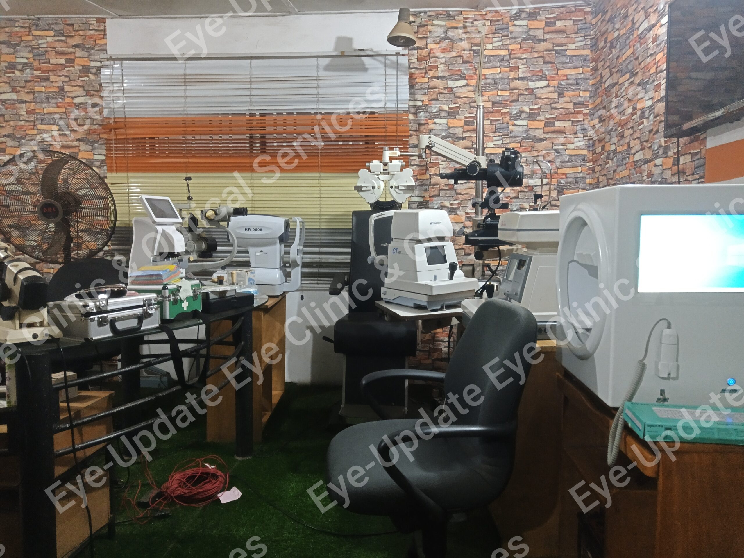
Dr. Dr. Steven E.N., Eyeupdate clinic & optical supplies, 01 Ajuwon junction, beside BPNL filling station, Ajuwon, off Elliot bus stop, Iju-Ishagah, Lagos. Tel: 08107531046, 08034971582.
Eye exam/test consists of series of tests that your eye doctor will perform in order to determine your ocular status.
An eye exam involves a series of tests to evaluate your vision and check for eye diseases/vision defects. Your eye doctor may use a variety of instruments, shine bright lights directly at your eyes and request that you look through an array of lenses. Each test during an eye exam evaluates a different aspect of your vision or eye health.
Why it’s done
An eye exam helps detect eye problems at their earliest stage — when they’re most treatable. Regular eye exams give your eye care professional a chance to help you correct or adapt to vision changes and provide you with tips on caring for your eyes.
When to have an eye exam
Several factors may determine how frequently you need an eye exam, including your age, health and risk of developing eye problems. General guidelines are as follows:
Children 3 years and younger
For children under 3, your pediatrician will likely look for the most common eye problems — lazy eye, crossed eyes or misaligned eyes. If there are eye concerns or symptoms, an examination is appropriate at that time regardless of age. Your child could undergo a more comprehensive eye exam between the ages of 3 and 5.
School-age children and adolescents
Have your child’s vision checked before he or she enters first grade. If your child has no symptoms of vision problems and no family history of vision problems, have his or her vision rechecked every one to two years. Otherwise, schedule eye exams based on the advice of your eye doctor.
Adults
In general, if you are healthy and you have no symptoms of vision problems, have your eyes checked on this schedule:
- Every five to 10 years in your 20s and 30s
- Every two to four years from 40 to 54
- Every one to three years from 55 to 64
- Every one to two years after age 65
Have your eyes checked more often if you:
- Wear glasses or contact lenses
- Have a family history of eye disease or loss of vision
- Have a chronic disease that puts you at greater risk of eye disease, such as diabetes
- Take medications that have serious eye side effects
-
How you prepare
There are three different types of eye specialists. Which specialist you choose may be a matter of personal preference or will depend on the nature of your eye problem.
- Ophthalmologists. Ophthalmologists are medical doctors who provide full eye care, such as giving you a complete eye exam, prescribing corrective lenses, diagnosing and treating complex eye diseases, and performing eye surgery.
- Optometrists. Optometrists provide many of the same services as ophthalmologists, such as evaluating your vision, prescribing corrective lenses, diagnosing common eye disorders and treating selected eye diseases with drugs. If you have a complex eye problem or need surgery, your doctor can refer you to an ophthalmologist.
- Opticians. Opticians fill prescriptions for eyeglasses, including assembling, fitting and selling them. Some opticians also sell contact lenses. Opticians do not provide eye health evaluations.
Bring your prescription eyewear
If you wear contact lenses or glasses, bring them to your appointment. Your eye doctor will want to make sure your prescription is the best one for you.
Other precautions
If your eyes are dilated as a part of your eye exam, you may want to bring sunglasses to wear after your eye exam is complete, as daylight or other bright lights may be uncomfortable or cause blurred vision. Also, consider having someone else drive you home.
What you can expect
Before the exam
If you’re seeing a new eye doctor or if you’re having your first eye exam, expect questions about your vision history. Part of the examination, such as taking your medical history and the initial eye test, may be performed by a clinical assistant or technician.
Your answers help your eye doctor understand your risk of eye disease and vision problems. Be prepared to give specific information, including:
- Are you having any eye problems now?
- Have you had any eye problems in the past?
- Do you wear glasses or contacts now? If so, are you satisfied with them?
- What health problems have you had in recent years?
- Were you born prematurely?
- Are you taking any medications?
- Do you have any allergies to medications, food or other substances?
- Have you ever had eye surgery?
- Does anyone in your family have eye problems, such as macular degeneration, glaucoma or retinal detachments?
- Do you or does anyone in your family have diabetes, high blood pressure, heart disease or any other health problems that can affect the whole body?
During the exam
An eye exam usually involves these steps:
- You’ll be asked about your medical history and any vision problems you might be experiencing.
- Your eye doctor measures your visual acuity to see if you need glasses or contact lenses to improve your vision.
- You’ll be given a numbing drop in your eyes. Then your eye pressure is measured. To make it easier for your doctor to examine the inside of your eye, he or she will likely dilate your eyes with eyedrops.
- After waiting for the dilating drops to take effect, your eye doctor checks the health of your eyes, possibly using several lights to evaluate the front of the eye and the inside of each eye.
Several different tests may be performed during the eye exam. The tests are designed to check your vision and to examine the appearance and function of all parts of your eyes.
After the exam
At the end of your eye exam, you and your doctor will discuss the results of all testing, including an assessment of your vision, your risk of eye disease and preventive measures you can take to protect your eyesight.
Different types of eye exams
Eye muscle test
This test evaluates the muscles that control eye movement. Your eye doctor watches your eye movements as you follow a moving object, such as a pen or small light, with your eyes. He or she looks for muscle weakness, poor control or poor coordination.
Visual acuity test
This test measures how clearly you see. Your doctor asks you to identify different letters of the alphabet printed on a chart (Snellen chart) or a screen positioned some distance away. The lines of type get smaller as you move down the chart. Each eye is tested separately. Your near vision also may be tested, using a card with letters similar to the distant eye chart. The card is held at reading distance.
Refraction assessment
Light waves are bent as they pass through your cornea and lens. If light rays don’t focus perfectly on the back of your eye, you have a refractive error. Having a refractive error may mean you need some form of correction, such as glasses, contact lenses or refractive surgery, to see as clearly as possible.
Assessment of your refractive error helps your doctor determine a lens prescription that will give you the sharpest, most comfortable vision. The assessment may also determine that you don’t need corrective lenses.
Your doctor may use a computerized refractor to estimate your prescription for glasses or contact lenses. Or he or she may use a technique called retinoscopy. In this procedure, the doctor shines a light into your eye and measures the refractive error by evaluating the movement of the light reflected by your retina back through your pupil.
Your eye doctor usually fine-tunes this refraction assessment by having you look through a masklike device that contains wheels of different lenses (phoropter). He or she asks you to judge which combination of lenses gives you the sharpest vision.
Visual field test (perimetry)
Your visual field is the full extent of what you can see to the sides without moving your eyes. The visual field test determines whether you have difficulty seeing in any areas of your overall field of vision. The different types of visual field tests include:
- Confrontation exam. Your eye doctor sits directly in front of you and asks you to cover one eye. You look straight ahead and tell the doctor each time you see his or her hand move into view.
- Manual testing, including tangent screen and Goldmann exams. You sit a short distance from a screen and focus on a target at its center. You tell the doctor when you can see an object move into your peripheral vision and when it disappears.
- Automated perimetry. As you look at a screen with blinking lights on it, you press a button each time you see a blink.
Using your responses to one or more of these tests, your eye doctor determines the fullness of your field of vision. If you aren’t able to see in certain areas, noting the pattern of your visual field loss may help your eye doctor diagnose your eye condition.
Color vision testing
You could have poor color vision and not even realize it. If you have difficulty distinguishing certain colors, your eye doctor may screen your vision for a color deficiency. To do this, your doctor shows you several multicolored dot-pattern tests.
If you have no color deficiency, you’ll be able to pick out numbers and shapes from within the dot patterns. If you do have a color deficiency, you’ll find it difficult to see certain patterns within the dots. Your doctor may use other tests, as well.
Slit-lamp examination
A slit lamp is a microscope that magnifies and illuminates the front of your eye with an intense line of light. Your doctor uses this device to examine the eyelids, lashes, cornea, iris, lens and fluid chamber between your cornea and iris.
Your doctor may use a dye, most commonly fluorescein (flooh-RES-een), to color the film of tears over your eye. This helps reveal any damaged cells on the front of your eye. Your tears wash the dye from the surface of your eye fairly quickly.
Retinal examination
A retinal examination — sometimes called ophthalmoscopy or funduscopy — allows your doctor to evaluate the back of your eye, including the retina, the optic disk and the underlying layer of blood vessels that nourish the retina (choroid). Usually before your doctor can see these structures, your pupils must be dilated with eyedrops that keep the pupil from getting smaller when your doctor shines light into the eye.
After administering eyedrops and giving them time to work, your eye doctor may use one or more of these techniques to view the back of your eye:
- Direct exam. Your eye doctor uses an ophthalmoscope to shine a beam of light through your pupil to see the back of the eye. Sometimes eyedrops aren’t necessary to dilate your eyes before this exam.
- Indirect exam. During this exam, you might lie down, recline in a chair or sit up. Your eye doctor examines the inside of the eye with the aid of a condensing lens and a bright light mounted on his or her forehead. This exam lets your doctor see the retina and other structures inside your eye in great detail and in three dimensions.
Screening for glaucoma
Tonometry measures the fluid pressure inside your eye (intraocular pressure). This is one test that helps your eye doctor detect glaucoma, a disease that damages the optic nerve.
Several methods to measure intraocular pressure are available, including:
- Applanation tonometry. This test measures the amount of force needed to temporarily flatten a part of your cornea. You’ll be given eyedrops with fluorescein, the same dye used in a regular slit-lamp examination. You’ll also receive eyedrops containing an anesthetic. Using the slit lamp, your doctor moves the tonometer to touch your cornea and determine the eye pressure. Because your eye is numbed, the test doesn’t hurt.
- Noncontact tonometry. This method uses a puff of air to estimate the pressure in your eye. No instruments touch your eye, so you won’t need an anesthetic. You’ll feel a momentary pulse of air on your eye, which can be startling.
If your eye pressure is higher than average or your optic nerve looks unusual, your doctor may use a pachometer. This instrument uses sound waves to measure the thickness of your cornea. The most common way of measuring corneal thickness is to put an anesthetic drop in your eye, then place a small probe in contact with the front surface of the eye. The measurement takes seconds.
You may need more-specialized tests, depending on your age, medical history and risk of developing eye disease.
Results
Normal results from an eye exam include:
- 20/20 vision
- Good peripheral vision
- Ability to distinguish various colors
- Normal-appearing structures of the external eye
- Absence of cataract, glaucoma or retinal disorders, such as macular degeneration
Your doctor may give you a prescription for corrective lenses. If your eye exam yields other abnormal results, your doctor will discuss with you next steps for further testing or for treating an underlying condition.
Leave a Reply
You must be logged in to post a comment.